
The inflammatory response of the immune system is a crucial function to control microbial infections. This response is facilitated by a highly inflammatory form of cell death called pyroptosis. To occur, pyroptosis depends on the activities of members of the mammalian caspase family of cysteinyl aspartate-specific proteases. Of these caspases, caspase-1 plays a pivotal role in this immune response by recognizing foreign danger signals and initiating a two-fold response.
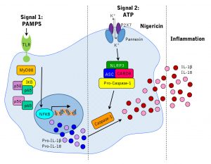
Casapse-1, initially known as interleukin converting enzyme (ICE), converts the important inflammatory cytokines interleukin 1β and interleukin 18 into their active forms. In addition, caspase-1 functions to mediate the lytic, programmed cell death of pyroptosis. Figure 1 illustrates how caspase-1 is activated by the NLRP3 inflammasome and converts IL-1β and IL-18 to be secreted.
Figure 1:
At ICT, apoptosis has always been a primary focus. However, pyroptosis is distinct from apoptotic cell death in that it results in plasma-membrane rupture and the release of pro-inflammatory cytokines. Cells that are infected by an intracellular pathogen will eventually swell, burst, and die during pyroptotic cell death. This attracts other immune cells to fight the infection, leading to overall tissue inflammation, and a clearance of the infection. This is in contrast to apoptotic cell death, which is non-inflammatory and characterized by the cells shrinking, membrane blebbing, and being phagocytosed by macrophages.
If you are studying the immune response or researching inflammation-related diseases such as asthma, rheumatoid arthritis, or multiple sclerosis, detecting pyroptosis through caspase-1 activity in cell culture can help you understand how your cells are dying.
ICT’s Pyroptosis/Casapse-1 Assay can quickly and easily assess pyroptotic cells and provides nigericin as a positive control for pyroptosis studies. This assay utilizes ICT’s popular fluorescent FLICA probe, which covalently binds with active caspase enzymes. Once FLICA is bound, it will retain the fluorescent signal within the cell, so pyroptotic cells will retain a higher concentration of FLICA and fluorescent brighter than negative cells. Cells can then be read using a flow cytometer, fluorescent plate reader, or fluorescence microscope.
Figure 2 shows a time-course response in Jurkat cells to nigericin-induced caspase-1 activation. ICT’s caspase-1 inhibitor reagent FAM-YVAD-FMK was used to monitor the caspase-1 induction response in Jurkat cells treated with Nigericin for various periods of time. The amount of caspase-1 activity detected directly correlated to the duration of the exposure period; the longer the cells were exposed to Nigericin, the larger the proportion of caspase-1 positive cells found in the sample.
Figure 2:
