Green Live/Dead Stain
A live-cell impermeant, green fluorescent DNA dye for viability, apoptosis and necrosis studies, and fixed cell nuclear counterstaining. Identifies necrotic or permeabilized cells. Analyze samples using a flow cytometer or fluorescence microscope.


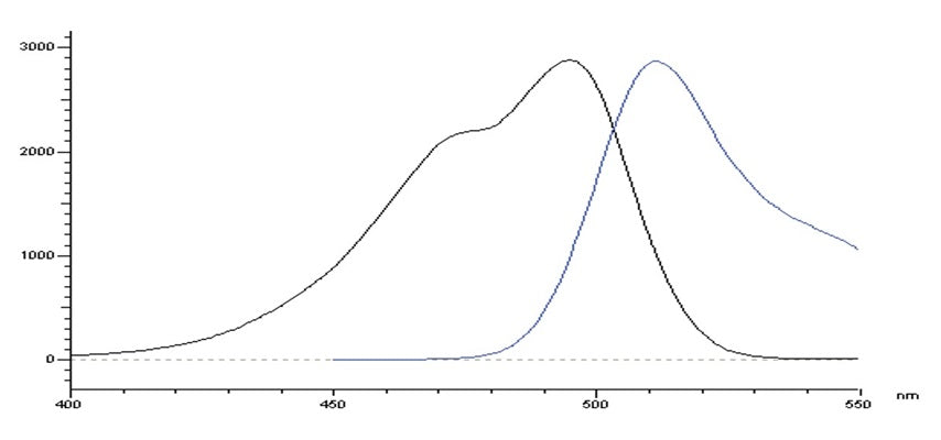
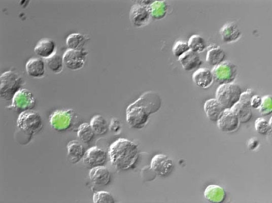
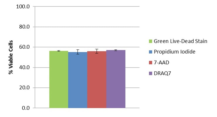
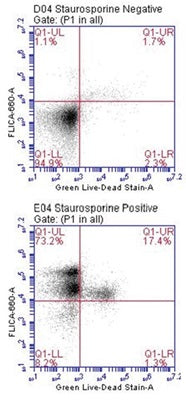
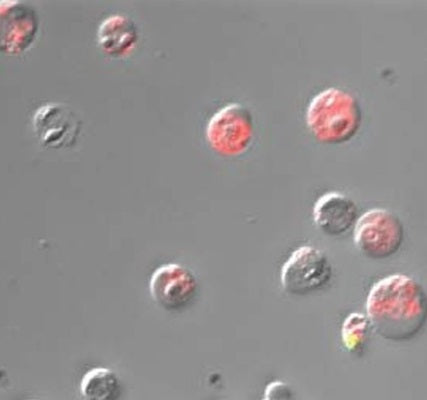
Green Live/Dead Stain (catalog #6342) is a vital dye that exhibits intact cell membrane exclusion properties analogous to the popular red fluorescing vital dyes, Propidium Iodide (PI), 7-amino- actinomycin D (7-AAD) and DRAQ7™. Like the red fluorescing dyes, Green Live/Dead Stain is excluded from intact, healthy cells due to its polar nature. In the presence of cells exhibiting compromised membrane integrity, Green Live/Dead Stain penetrates the cell and nuclear membrane barriers and intercalates tightly to DNA in a manner analogous to PI and 7-AAD. When bound to DNA, it acquires a greatly enhanced fluorescence potential (>2000X) in the green emission range. These important vital dye properties enable Green Live/Dead Stain to be used in flow cytometry-based protocols to assess the percentage of late apoptotic, necrotic, and membrane-compromised cells within a sample cell population. When bound to nucleic acids, the maximum absorption of Green Live/Dead Stain is 495 nm and the maximum emission is 512 nm (Figure 1). Cells can be viewed through a fluorescence microscope (Figures 2 & 5) or analyzed with a flow cytometer (Figure 4). Green Live/Dead Stain is provided as a 500 µM concentrated stock solution dissolved in DMSO. For flow cytometry applications, a staining concentration of 50 nM is recommended. Therefore, using sample sizes of 0.5 mL, a single 50 µL vial (catalog #6342) provides enough reagent for 1000 tests. For fluorescence microscopy applications, a usage concentration of 0.5 µM is suggested. In this way, a vial is sufficient for 100 tests (0.45 mL sample sizes). Green Live/ Dead Stain can be used with ICT’s red FLICA® 660 caspase inhibitor reagents (e.g., catalog #9120) to identify four populations of cells: living; early apoptotic; late apoptotic; and necrotic (Figures 4 & 5).
- Each 50 µL vial is sufficient for 1000 flow cytometry tests.
- Spin thawed vial briefly in a microcentrifuge to remove any reagent that may have become trapped in the cap.
- Dilute Green Live/Dead Stain concentrated stock solution (500 µM) 1:100 in PBS to prepare a 5000 nM working solution. For example, add 10 µL stock concentrate to 990 µL PBS. Prepare only what is needed for the experiment. Working solution should be used immediately, and any remaining diluted solution should be discarded.
- Spike samples with a 1:100 dilution of the 5000 nM working solution. For example, spike 0.495 mL samples with 5 µL working solution.
- Incubate samples for ~10 minutes at room temperature, protected from light.
- Analyze with a flow cytometer using a blue laser at 488 nm and a 530/30 (FL1) emission filter setting, or similar.
- Each 50 µL vial is sufficient for 100 microtiter plate wells.
- Spin thawed vial briefly in a microcentrifuge to remove any reagent that may have become trapped in the cap.
- Dilute Green Live/Dead Stain concentrated stock solution (500 µM) 1:100 in PBS to prepare a 5000 nM working solution. For example, add 10 µL stock concentrate to 990 µL PBS. Prepare only what is needed for the experiment. Working solution should be used immediately, and any remaining diluted solution should be discarded.
- Spike samples with a 1:10 dilution of the 5000 nM working solution. For example, spike .45 mL samples with 50 µL working solution.
- Incubate samples for ~10 minutes at room temperature, protected from light.
- When bound to DNA, the peak absorption of Green Live/Dead Stain is 495 nm and the maximum emission is 512 nm. Visualize with a fluorescence microscope using optical filters that best approximate these settings.
Product Specific References
| PMID | Publication |
| 38008083 | Germain, A., et al. 2023. Co-culture device for in vitro high throughput analysis of cancer associated fibroblast and cancer cell interactions. Oncology. |
| 35225960 | Halloran, Daniel R., D, et al. 2022. Differentiation of Cells Isolated from Human Femoral Heads into Functional Osteoclasts. Journal of Developmental Biology, 6. |