
If you’re first branching into cellular analysis, the amount of assay options for detecting apoptosis can be overwhelming. Here, we’ll explore one method of apoptosis detection: Annexin V-Fluorescein.
What are annexins?
Annexins are cellular proteins that meet two basic criteria:
- They are capable of binding negatively charged phospholipids in a calcium dependent manner.
- They contain a 70 amino acid sequence called an annexin repeat.
Found in eukaryotic cells, annexins play an important role in providing a cell membrane scaffold that is relevant to changes in the physical shape of the cell. As such, they are involved in a variety of cellular processes such as exocytosis, endocytosis, trafficking and organization of vesicles, and calcium ion channel formation. The Annexin V protein is a member of a calcium and phospholipid binding family of proteins with anticoagulant properties, and studies show that it may play a role in the inhibition of blood coagulation.
Apoptosis detection
So, how does Annexin V relate to apoptosis? It is thought that Annexin V possesses its ability to inhibit blood coagulation by competing for phosphatidylserine (PS) binding sites with prothrombin, a protein in blood plasma that contributes to blood clotting appropriately. In healthy cells, PS is kept on the inner-leaflet, or cytosolic, side of the cell membrane. In apoptotic cells, the cell quickly becomes asymmetric, and PS translocates to be exposed on the surface of the cell. Using a recombinant fluorescein-conjugated Annexin V with Propidium Iodide (PI) allows you to distinguish two populations of dying cells from viable cells. Apoptotic cells with an intact membrane and the surface-exposed PS will stain green for Annexin V-Fluorescein. Cells that are in later stage apoptosis that have lost membrane integrity with stain both green and red for Annexin V-Fluorescin and PI. Live, non-apoptotic cells will not stain positive for either treatment.
From the bench
In this experiment, Jurkat cells were treated with a negative control (top) or staurosporine (bottom) for 4 hours, washed, and stained with ICT’s Bovine Annexin V-Fluorescein Assay for 15 minutes. Cells were read on an Accuri C6 flow cytometer. Treatment with the negative control induced Annexin V-Fluorescin labeling in only 17.6% of the cell population (UR + LR = 13.5% + 4.1% left), whereas treatment with staurosporine induced Annexin V-Fluorescin labeling in 98.8% of the experimental cells (UR + LR = 15.3% + 83.5% right). Treatment with staurosporine did not significantly increase the proportion of dually stained cells (Annexin V-Fluorescin(+)/PI(+), UR) at 4 hours. Data courtesy of Mrs. Tracy Hansen.

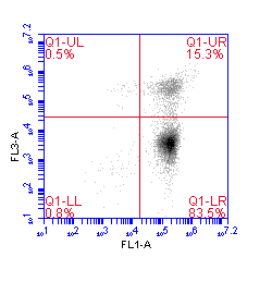
Annexin V-Fluorescein, FL-1 Negative Control, Non-Induced
Products
Annexin V-Fluorescein kits are available for a variety of species, including bovine, canine, chicken, equine, and swine. These kits are useful for those looking for a fast and easy assay to assess apoptosis in cell culture. If you are looking for a more sophisticated apoptosis detection assay that assesses the apoptotic process rather than the effects of apoptosis (as with the Annexin V assays), we would encourage you to explore ICT’s line of FLICA caspase assays.