Basic Calcein AM Cell Viability Kit
This kit allows researchers to easily and simultaneously differentiate between live and dead cells within a single sample. Samples can be analyzed using a flow cytometer, fluorescence plate reader, or fluorescent microscope.


Assessment of cell viability is a critical step during the evaluation of novel drug treatments and therapies for potential cytotoxic properties. With cell viability assessment playing a central role in countless research and environmental safety studies, there is an ever present need for simple, straightforward analysis methods capable of distinguishing between live and dead cells. The Basic Calcein AM Cell Viability kit developed by ImmunoChemistry Technologies (ICT) allows for easy and simultaneous differentiation of live and dead cells within a single sample.
To use Calcein AM, simply add the reagent directly to the cell sample, incubate, and analyze (no wash steps necessary). Calcein AM is a membrane permeant, fluorogenic reagent widely recognized for its utility in assessing the relative cell viability status of different cell populations. Calcein AM’s overall hydrophobic nature allows it to readily traverse the lipid bilayer structure of the cell membrane in a concentration gradient-dependent manner. Once inside the cell, the hydrophobic and non-fluorescent Calcein AM is quickly hydrolyzed by intracellular esterases that are active in live cells. This leads to the cleavage and removal of two non-polar acetoxymethyl ester (AM) groups. Once the AM groups have been cleaved, the resulting polar (hydrophilic) and now fluorescence-capable Calcein dye molecule is efficiently retained within the confines of the cell membrane. Polar dye molecules will naturally be excluded from passive diffusion back out of the cell again due to the hydrophobic lipid bilayer composition of the cell membrane. Dead cells lack active esterases and do not cleave Calcein AM.
The large quantum yield of Calcein dyes enables them to be readily detected within widely used applications such as flow cytometers, fluorescence plate readers, and fluorescence microscopes. The degree of fluorescence correlates with relative cell viability status. For microscopy usage, Hoechst 33342 is included with the kit to concurrently label nuclei after labeling with Calcein. Calcein optimally excites at 494 nm with maximal emission at 517 nm. Hoechst 33342 can be seen using a UV-filter with excitation at 365 nm and emission at 480 nm.
- Prepare samples and controls.
- Dilute 10X Cellular Assay Buffer 1:10 with diH2O.
- Reconstitute Calcein AM with 50 µL DMSO to prepare 2 mM stock solution.
- Dilute 2 mM Calcein AM stock solution 1:5 by adding 200 µL diH2O or PBS, forming a 400 µM stock solution.
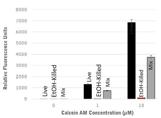
- Stain with Calcein AM at a concentration between 1-10 µM. The ideal staining concentration can vary based on cell line, application, etc., and should be determined by the end user. The recommended sample size is 0.4 mL.
- Add Calcein AM to each sample and mix gently. For example, to stain a 0.4 mL sized sample with 10 µM Calcein AM, add 10 µL of the 400 µM stock solution.
- Incubate approximately 1 hour.
- Analzye with a flow cytometer, fluorescence microscope, or fluorescence plate reader. Calcein AM excites at 494 nm and emits at 520 nm.