Green Fluorescent Cathepsin B Assay
The quenched substrate Rhodamine 110-(RR)2, fluoresces green upon cleavage by active cathepsins, allowing the monitoring of intracellular cathepsin activity in vitro using live whole cells. Analyze using flow cytometry or fluorescence microscopy.


Our Green Fluorescent Cathepsin B Assay enables researchers to quantitate and monitor intracellular cathepsin activity over time in vitro. The Rhodamine 110 Cathepsin B substrate reagent is a non-cytotoxic and membrane permeant substrate that fluoresces green upon cleavage by active cathepsin enzymes.
Cathepsins are a group of protease enzymes that were originally identified in lysosomes. Cathepsins are classified based on the key catalytic group present in their active site, and are categorized as aspartic, serine, or cysteine proteases. Cathepsins D and E are aspartic proteases, cathepsins A and G are serine proteases, and cathepsins B, C, F, H, K, L, O, S, V, W, and X are cysteine proteases. Initially synthesized as inactive zymogens, they are post-translationally processed into their active configurations after passing through the endoplasmic reticulum and subsequent incorporation into the acidic environment of the lysosomes. These enzymes exist in their processed form as disulfide-linked heavy and light chain subunits with molecular weights ranging from 20-35 kDa. Cathepsin C is the noted exception, existing as an oligomeric enzyme with a MW ~200 kDa.
Cathepsin B is thought to play important roles in many cellular functions, including intracellular protein degradation, antigen processing and presentation, and proenzyme processing, to name a few. Elevated cathepsin enzyme activity in serum or the extracellular matrix often signifies a number of gross pathological conditions. Cathepsin-mediated diseases include: neurodegenerative diseases such as Alzheimer’s; autoimmune related diseases like arthritis; and the accelerated breakdown of bone structure seen with osteoporosis. Up-regulated cathepsin B activity has also been linked to several types of cancer. These include cancer of the colon, pancreas, ovaries, breast, lung, and skin (melanoma).
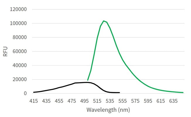
Rhodamine 110 Cathepsin B substrate utilizes the photostable green fluorophore, rhodamine 110. Our Rhodamine 110 Cathepsin B substrate is comprised of rhodamine 110 coupled to two copies of the amino acid sequence, arginine-arginine (RR), which is the preferential target sequence for cathepsin B. When bi-substituted via amide linkage to two cathepsin B target peptide sequences, rhodamine 110 is nonfluorescent. Following enzymatic cleavage at one or both arginine (R) amide linkage sites, the mono and non-substituted rhodamine 110 fluorophores generate green fluorescence when excited at 500 nm.
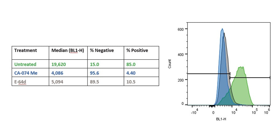
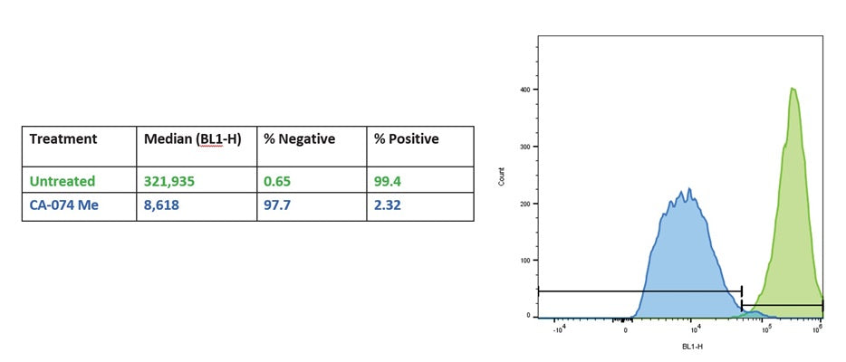
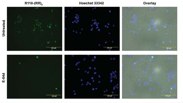
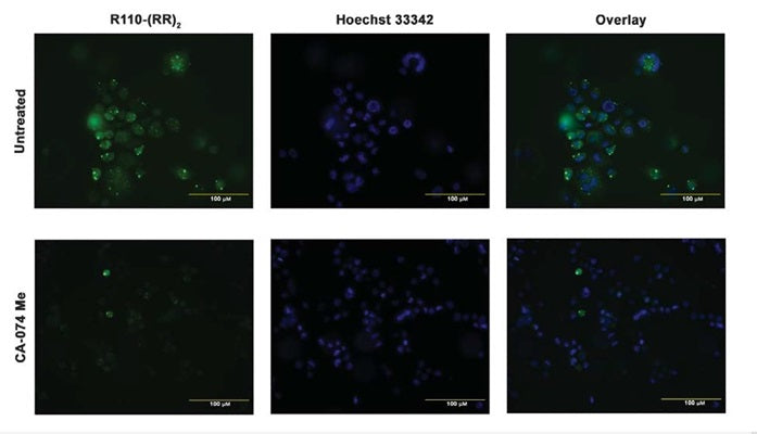
To use the Green Cathepsin B Assay, simply add the Rhodamine 110 Cathepsin B substrate [R110-(RR)2] directly to the cell culture media (or 1X Cellular Assay Buffer), incubate, and analyze. Because R110-(RR)2 is cell-permeant, it easily penetrates the cell membrane and the membranes of the internal cellular organelles - no lysis or permeabilization steps are required. R110-(RR)2 will enter the cell in a non-fluorescent state. If cathepsin enzymes are active, they will cleave off the two arginine-arginine cathepsin B targeting sequences and allow the rhodamine 110 fluorophore to become fluorescent upon excitation. By varying the duration and concentration of exposure to the R110-(RR)2 substrate, a picture can be obtained of the relative abundance and intracellular location of cathepsin enzymatic activity. Positive cells will fluoresce green, while negative cells will exhibit very low levels of background green fluorescence. There is no interference from pro-cathepsins forms of the enzymes. If the treatment or experimental condition stimulates cathepsin activity, cells containing elevated levels of cathepsin activity will appear brighter green than cells with lower levels of cathepsin activity.
R110-(RR)2 has an optimal excitation of 500 nm and emission of 525 nm. Hoechst 33342 is included with the kit to concurrently label nuclei after labeling with R110-(RR)2. Hoechst 33342 is revealed under a microscope using a UV-filter with excitation at 365 nm and emission at 480 nm. Cells can be easily analyzed by flow cytometry or fluorescence microscopy.
- Dilute 10X Cellular Assay Buffer 1:10 with diH2O.
- Prepare samples and controls in fresh cell culture medium or 1X Cellular Assay Buffer.
- Reconstitute R110-(RR)2 with 50 µL DMSO.
- Dilute R110-(RR)2 1:5 by adding 200 µL PBS.
- Add diluted R110-(RR)2 to each sample at 1:50 (e.g., add 10 µL of diluted R110-(RR)2 to 490 µL of sample).
- Incubate while protected from light.
- If desired, label with additional stains, such as Hoechst 33342, DAPI, or an antibody.
- Analyze with a flow cytometer or fluorescence microscope. R110-(RR)2 excites at 500 nm and emits at 525 nm.